Persistent right aortic arch and associated anatomical anomalies in dogs (Canis lupus familiaris)
Keywords:
Right aortic arch;, vascular ring, esophagus, dogAbstract
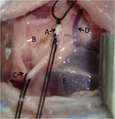
Persistent fourth right aortic arch is considered one of the most frequent anatomical abnormalities in vascular ring formation. Structure of this type of ring is formed by the aorta originated from the fourth right aortic arch, pulmonary trunk and left arteriosus ligament. Cases described in this study correspond to this type of anomaly, the patients were diagnosed and then underwent surgical treatment to correct the vascular defect. During surgery, the vascular ring observed, which compressed esophagus and trachea. In addition, megaesophagus (cranial to the heart), and presence of a left azygos vein and the formation of connective tissue bands around the esophagus were observed as associated anatomical abnormalities. Left azygos vein was not sectioned because it did not cause esophageal compression in any of cases. These findings coincide with that reported in the literature and reinforce the theory that this type of vascular ring is the most frequent.
Downloads
Published
Issue
Section
License
Gaceta de Ciencias Veterinarias se apega al modelo Open Access, por ello no se exige suscripción, registro o tarifa de acceso a los usuarios o instituciones. Los usuarios pueden leer, descargar, copiar, distribuir, imprimir y compartir los textos completos inmediatamente después de publicados, se exige no hacer uso comercial de las publicaciones. Para la reproducción parcial o total de los trabajos o contenidos publicados, se exige reconocer los derechos intelectuales de los autores y además, hacer referencia a esta revista. La publicación de artículos se hace sin cargo para los autores. Los trabajos pueden consultarse y descargarse libremente, y de manera gratuita, en extenso en versión digital, desde su enlace Web institucional. Los textos publicados son propiedad intelectual de sus autores. Las ideas, opiniones y conceptos expuestos en los trabajos publicados en la revista representan la opinión de sus autores, por lo tanto, son estos los responsables exclusivos de los mismos.